About this case & sources
This published patient developed acute pericardial effusion and tamponade after an inferior vena cava filter was removed. This recording was acquired before drainage. This lesson identifies fluid; the outline alone cannot establish tamponade or its severity.
Zhang, Wang & Liu (2021), Additional file 1, CC BY 4.0. A short apical four-chamber cine was cropped to remove peripheral identifiers and display text. The moving contour is an inset educational guide to the visible apical and image-left fluid, not a measured segmentation. Chamber contours are inset orientation guides to visible regions; the valve pointers indicate the atrioventricular junctions, not individual leaflet borders. Clinical review is pending.
Published case & original videos ↗CC BY 4.0 ↗Echo interpretation reference ↗ASE examination reference ↗Credits & image preparation ↗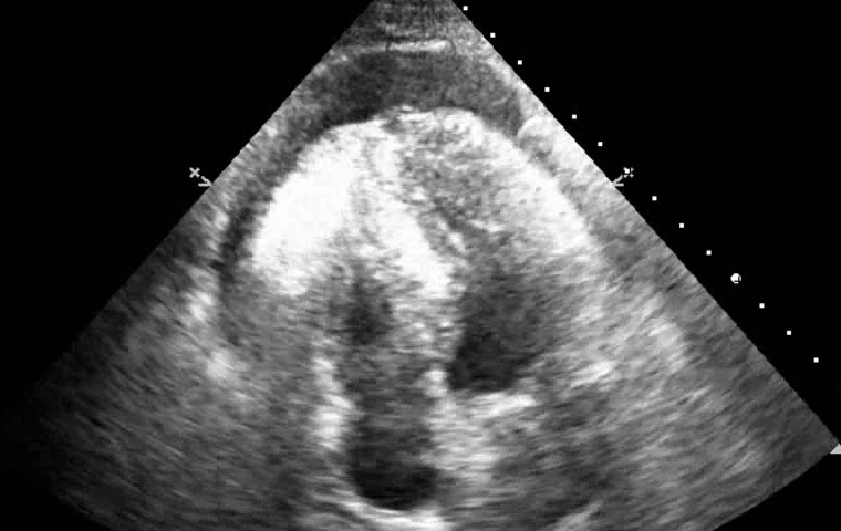 Pericardial effusion